Stage Function In Microscope

The Stage Function in Microscopes: A Comprehensive Exploration
Microscopes are indispensable tools in scientific research, education, and industry, enabling the visualization of structures and organisms invisible to the naked eye. Among its critical components, the stage plays a pivotal role in sample positioning, stability, and observation accuracy. This article delives into the functions, types, and advancements of microscope stages, combining historical context, technical insights, and practical applications.
Historical Evolution of Microscope Stages
The microscope stage has evolved significantly since the invention of the first compound microscope in the late 16th century. Early designs featured simple, fixed platforms made of wood or metal, offering limited adjustability. By the 19th century, mechanical stages emerged, incorporating precise movement controls. The 20th century introduced motorized and automated stages, revolutionizing fields like cell biology and materials science.
Core Functions of the Microscope Stage
1. Sample Support and Stability
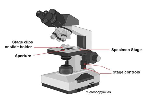
The primary function of the stage is to hold the specimen securely during observation. Modern stages are engineered with materials like aluminum or stainless steel, ensuring durability and resistance to environmental factors.
2. Precise Positioning
Mechanical stages allow for fine adjustments in the x, y, and z axes. This precision is essential for mapping large samples or focusing on specific areas. For example, in histopathology, pathologists rely on stage movement to examine tissue sections systematically.
3. Compatibility with Accessories
Stages often integrate with accessories like:
- Condensers: Enhance light focusing for brighter images.
- Polarizers: Enable polarized light microscopy for birefringent materials.
- Heaters/Coolers: Maintain sample temperature in live-cell imaging.
| Accessory | Function | Application |
|---|---|---|
| Polarizer | Filters light waves | Mineralogy, polymer analysis |
| Heater Stage | Controls temperature | Live-cell imaging, phase transitions |

Types of Microscope Stages
Mechanical Stages
These stages feature manual controls (knobs or levers) for x-y movement. They are cost-effective and widely used in educational settings. However, they require skill to operate without displacing the sample.
Motorized Stages
Powered by stepper or servo motors, these stages offer automated movement, programmable coordinates, and repeatability. They are essential in high-throughput imaging and mapping applications.
Inverted Stages
Used in inverted microscopes, these stages position the sample below the objective lens. This design facilitates imaging of cell cultures in multiwell plates or heavy specimens.
Specialized Stages
- Rotation Stages: Allow 360° sample rotation for crystallography.
- Tilt Stages: Enable angular adjustments for 3D imaging.
- Flow-Through Stages: Support liquid samples in fluid dynamics studies.
Technological Advancements in Stage Design
Automation and Robotics
Modern stages integrate with software for automated workflows. For instance, AI-driven systems can identify regions of interest (ROIs) and map entire samples autonomously.
Nanopositioning Stages
These stages achieve sub-nanometer precision using piezoelectric actuators. They are vital in nanoscale research, such as imaging quantum dots or graphene layers.
"Nanopositioning stages have transformed materials science by enabling atomic-level resolution," states Prof. Raj Patel, a nanotechnologist at MIT.
Environmental Control Stages
Stages with integrated chambers maintain humidity, gas composition, or temperature. This is crucial for observing living organisms or chemical reactions in real time.
Practical Applications Across Disciplines
Biomedical Research
In cancer research, motorized stages map tumor tissues to identify metastasis patterns. In neuroscience, they track neuronal activity in live brain slices.
Materials Science
Stages with polarizers analyze polymer structures, while heated stages study phase transitions in alloys.
Forensics
Forensic labs use stages to examine fibers, hairs, or gunshot residues under high magnification.
Challenges and Limitations
While stages are indispensable, they face challenges:
- Mechanical Drift: Thermal expansion or wear can cause gradual misalignment.
- Load Capacity: Heavy samples may exceed stage weight limits.
- Cost: Advanced stages (e.g., nanopositioners) are expensive, limiting accessibility.
Future Trends in Stage Technology
Emerging trends include:
1. AI Integration: Predictive algorithms optimize stage movement for faster imaging.
2. Hybrid Stages: Combine mechanical and piezoelectric systems for versatility.
3. Miniaturization: Compact stages for portable microscopes in field research.
What is the difference between a mechanical and motorized stage?
+Mechanical stages rely on manual adjustments, while motorized stages use motors for automated, precise movement. Motorized stages are faster and repeatable but more expensive.
Can microscope stages be used for live-cell imaging?
+Yes, stages with environmental control (e.g., temperature, CO₂ regulation) are designed for live-cell imaging, ensuring cell viability during observation.
How do nanopositioning stages achieve sub-nanometer precision?
+They use piezoelectric materials that expand or contract in response to voltage, enabling ultra-fine movements without mechanical parts.
Conclusion
The microscope stage, often overlooked, is a cornerstone of effective microscopy. From its humble beginnings to today’s high-tech designs, it exemplifies the intersection of engineering and biology. As technology advances, stages will continue to unlock new frontiers in visualization, shaping discoveries across disciplines.
Final Thought: Whether mapping tissues or analyzing nanomaterials, the stage’s role in microscopy is as foundational as the lens itself—a silent enabler of scientific progress.

